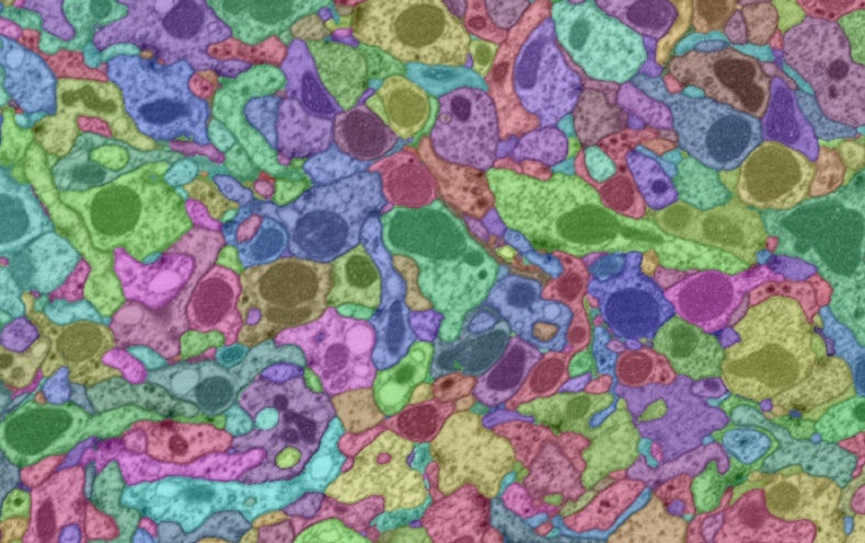
The brain of a common fruit fly (Drosophila melanogaster) is no larger than a poppy seed, but the miniscule piece of tissue holds tens of thousands of neurons joined by tens of millions of synapses. Although it is no rival to the human brain, which contains more than 80 billion neurons, the fly organ is still an invaluable tool for investigating the neural circuity underlying some of the basic behaviors of more complex animals. Using fruit flies, scientists have now constructed the largest brain map to date.
The dream of creating a fruit fly connectome—a wiring diagram of neurons and synapses—has been nurtured for more than a decade, says Gerry Rubin, executive director of the Howard Hughes Medical Institute’s Janelia Research Campus. Rubin and his colleagues in Janelia’s FlyEM group have now taken a big step toward making this goal a reality: They have completed a connectome of a region that covers roughly a third of the fly brain and includes areas involved in key functions, such as learning, smell and spatial navigation. The data set, which has been dubbed the hemibrain, contains more than 20,000 neurons and approximately 20 million synaptic connections. Today the team posted the data on Janelia’s Web site, along with an accompanying manuscript on the preprint server bioRxiv.
Rubin likens a connectome to Google Maps. In the same way that you use the navigation tool to search for a route from New York City to San Francisco, a connectome can reveal the pathways connecting neuron A and neuron B. Researchers have imaged the entire fruit fly brain in detail in the past but at lower resolutions. The only complete connectome to date is that of the roundworm Caenorhabditis elegans, which contains a mere 302 neurons. “I think the really big impact of this data release is going to be showing people the kind of insights you can get when you have a complete connectome of an animal that has interesting behaviors,” Rubin says.
This feat required the work of dozens of researchers from various disciplines, including biology, computer science and physics. It also involved a number of painstaking steps: slicing the samples, imaging them using focused ion beam scanning electron microscopy (FIB-SEM)—a technique that utilizes charged particles to slowly chip away at a chunk of tissue—and identifying and labeling neurons and synapses.
That last step was carried out by combining machine learning algorithms from Google with a team of proofreaders at Janelia. According to Rubin, the partnership with Google was facilitated by Viren Jain, a research scientist at the tech company who was formerly at Janelia. Jain and his colleagues were looking for a data set to test Google’s algorithms for imaging segmentation—a process that involves identifying the components of an image and labeling them. Janelia, on the other hand, was seeking help to analyze the massive trove of data generated by imaging the fruit fly brain.
“The Drosophila [neuroscience] community will profit from this connectome data enormously,” says Alexander Borst, a neuroscientist at the Max Planck Institute for Neurobiology in Martinsried, Germany. Borst, who is not a part of FlyEM but is one of Rubin’s collaborators, notes that there are some limitations to connectome data. While such information can save researchers a lot of time because it spares them from doing painstaking experiments in order to find the synaptic connections to their neuron of interest, “by itself, [a connectome] does not solve the puzzle of how the brain controls behavior,” he says. “It rather provides a data set, like a telephone book, where one can look up the phone number.”
The fruit fly brain itself also has some limitations. While the insects can be used to study functions such as navigation and olfaction, animals such as rodents or primates possess more behaviors that model our own. For this reason, scientists are also hoping to build a connectome of the mouse brain. And an advisory group to the director of the National Institutes of Health recently identified that plan as a “transformative project” in its BRAIN Initiative 2.0 report.There are some challenges, however. Not only are mouse brains much bigger than fly brains, there is also more variability between them. Unlike worms and flies, the brains of mice—and humans—are more malleable to experience, says Sebastian Seung, a neuroscientist at Princeton University. (Seung is not part of FlyEM but is involved in FlyWire, a project that has aimed to build a fly connectome with the help of crowdsourcing.)
The FlyEM group is now working on constructing a circuit diagram of the fruit fly’s full nervous system—which consists of the entire brain and nerve cord, a spinal cordlike structure in the insect. Stephen Plaza, the leader of the FlyEM project, says that he and his colleagues expect to complete this project within the next two years. In the meantime, he adds, the group plans to continue to assess the hemibrain data and update them if users pinpoint any mislabeled neurons of missed connections. “We don’t expect that this is the final chapter by any means,” Plaza says.
"brain" - Google News
January 22, 2020 at 08:00AM
https://ift.tt/30JETXG
Largest Brain Wiring Diagram to Date Is Published - Scientific American
"brain" - Google News
https://ift.tt/2W2PMS9
Shoes Man Tutorial
Pos News Update
Meme Update
Korean Entertainment News
Japan News Update
No comments:
Post a Comment